What is Gram Staining? Gram staining is a common technique used to differentiate two large groups of bacteria based on their different cell wall constituents. The Gram stain procedure distinguishes between Gram positive and Gram negative groups by coloring these cells red or violet. Gram positive bacteria stain violet due to the presence of a thick layer of peptidoglycan in their cell walls, which retains the crystal violet these cells are stained with. Alternatively, Gram negative bacteria stain red, which is attributed to a thinner peptidoglycan wall, which does not retain the crystal violet during the decoloring process. How Does Gram Staining Work? Gram staining involves three processes: staining with a water-soluble dye called crystal violet, decolorization, and counterstaining, usually with safanin. Due to differences in the thickness of a peptidoglycan layer in the cell membrane between Gram positive and Gram negative bacteria, Gram positive bacteria (with a thicker pept...
Granulocytes Are phagocytes, which have the ability to ingest viruses, bacteria and other parasites. They have visible granules or grains in their cytoplasm and have large elongated or lobed nuclei. The diameter of cell measures approximately from (12 – 20) μ, and their nucleoli cannot be seen. They account for approximately 60% of our WBCs. The sub-types of granulocytes are: neutrophil, basophil and eosinophil. Neutrophils are a part of the innate immune system and an essential line of defense against bacteria. The shape of nucleus is like a “U” or a curled rod prior to segmentation. They are also known as “band neutrophils”. The diameter usually ranges between (10–18)μ. The cytoplasm is moderate to abundant with a few non-specific granules. Neutrophils account for approximately (1%–3%) of the peripheral WBCs. The diameter of a segmented neutrophil cell usually ranges between (9–16)μ. They have a multi-lobed nucleus (three o...
Introduction Widal Test is an agglutination test which detects the presence of serum agglutinins (H and O) in patients serum with typhoid and paratyphoid fever. When facilities for culturing are not available, the Widal test is the reliable and can be of value in the diagnosis of typhoid fevers in endemic areas. It was developed by Georges Ferdinand Widal in 1896 The patient’s serum is tested for O and H antibodies (agglutinins) against the following antigen suspensions (usually stained suspensions): S. Typhi H antigen suspension, d S. Typhi 0 antigen suspension, 9, 12 S. Paratyphi A 0 antigen suspension, 1, 2, 12 S. Paratyphi A H antigen suspension, a S. Paratyphi B 0 antigen suspension, 1, 4, 5, 12 S. Paratyphi B H antigen suspension, b, phase 1 S. Paratyphi C 0 antigen suspension, 6, 7 S. Paratyphi C H antigen suspension, c, phase 1 Salmonella antibody starts appearing in serum at the end of first week and rise sharply during the 3rd week of endemic fever. In acute typhoid fev...
This post describes the use of disinfectants for routine laboratory decontamination of surfaces and equipment. Definitions: Disinfectants : Disinfectants are substances that are applied to non-living objects to destroy microorganisms that are living on the objects. Microorganisms : A microorganism or microbe is a microscopic organism that comprises either a single cell (unicellular), cell clusters, or multicellular relatively complex organisms. Antiseptic : Any substance that inhibits the growth and reproduction of microorganisms. Generally includes only those that are used on living objects (as opposed to disinfectants) and aren’t transported by the lymphatic system to destroy bacteria in the body (as opposed to antibiotics ). Decontamination – A process that removes the total burden of all classes of microorganisms, usually using chemicals, heat, and/or pressure. Disinfection – A process that reduces microbial burden on a surface or object. Inactivation – The pro...
Coagulase Test- Principle, Procedure, Types, Result, Uses Coagulase Test Definition A coagulase test is a biochemical test that is used to differentiate Staphylococcus aureus from other Staphylococci species like S. epidermidis and S. saprophyticus on the basis of the ability to produce the coagulase enzyme. The coagulase test is an important test that differentiates the species of the genus Staphylococci into two groups; Coagulase positive Staphylococci and Coagulase Negative Staphylococci. The coagulase enzyme acts as a virulence factor in some organisms as it interacts with the fibrinogen present on the host’s cell surface. Organisms with coagulase usually have a protective barrier around themselves, increasing their pathogenicity and resistance against the immune system. Coagulase is of two types; free coagulase and bound coagulase, each of which is detected by different methods. The bound coagulase is called the clumping factor and is detected rapidly by a slide test. The free c...
Definition Urinary tract infection (UTI) is the active infection in any part of urinary tract beyond distal urethra which is normally bacteriologically sterile. Causative Agents A large number of organisms gaining access to urinary tract are capable of causing UTI (Table 1). These organisms may reach the urinary tract through ascending route or by haematogenous route. Table 1 : Causative agents of UTI Factors Predisposing to UTI Various bacterial attributes and host factors favour urinary tract infections. Mechanical factors are important. Anything that disrupts normal urine flow or complete emptying of the bladder, or facilitates access of organisms to bladder, will predispose an individual to UTI. Clinical Specimen: Collection and Transportation Mid-stream sample of urine is the ideal specimen for the diagnosis of UTI. First part of the urine washes away the surface commensals from the distal urethra and hence the midstream specimen indicates actual bacteriological pi...
What is an autoimmune disease? An autoimmune disease is a condition in which your immune system mistakenly attacks your body tissues and cells as foreign and launches an inappropriate immune response. The immune system normally guards against germs like bacteria and viruses. When it senses these foreign invaders, it sends out an army of fighter cells to attack them. Normally, the immune system can tell the difference between foreign cells and your own cells. In an autoimmune disease, the immune system mistakes part of your body, like your joints or skin, as foreign. It releases proteins called autoantibodies that attack healthy cells. Some autoimmune diseases target only one organ. Type 1 diabetes damages the pancreas. Other diseases, like systemic lupus erythematosus (SLE), affect the whole body. Why does the immune syste attack the body? Doctors don’t know exactly what causes the immune-system misfire. Yet some people are more likely to get an autoimmune disease than others. Ac...
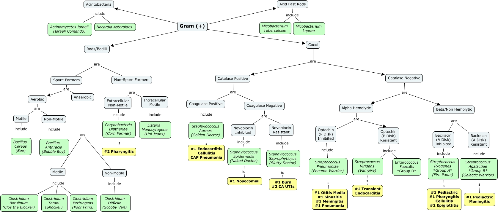
Morphological classification of medical important bacteria Morphologically bacteria can resemble: Cocci (Singular: coccus) Bacilli / rods (Singular: rod, bacillus) Vibrios (Singular: vibrio) Spirilla (Singular: spirillum) Spirochaetes (Singular: spirochaet Cocci : These are round or oval bacteria measuring about 0.5–1.0 m in diameter. When multiplying, cocci may form pairs, chains, or irregular groups: cocci in pairs are called diplococci, e.g. meningococci and gonococci. cocci in chains are called streptococci, e.g. Streptococcus pyogenes. cocci in irregular groups are called staphylococci, e.g. Staphylococcus aureus. Gram reaction : Staphylococci and streptococci are Gram positive, whereas diplococci can be Gram positive or Gram negative. Rods (bacilli): These are stick-like bacteria with rounded, tapered (fusiform), square, or swollen ends. They measure 1–10 µ m in length by 0.3–1.0 µ m in width. The short rods with rounded ends are often called coccobacilli. When mul...


Comments
Post a Comment